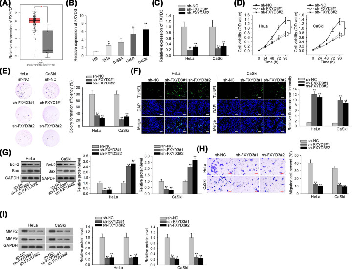
Figure 1. Overexpressed FXYD3 contributes to the progression of CC.
(A) FXYD3 expression level in CC tissues was identified by GEPIA2 database. (B) The expression level of FXYD3 in CC cell lines was assessed by RT-qPCR analysis. (C) RT-qPCR was applied to estimate the transfection efficiency of FXYD3 specific shRNAs. (D and E) CCK-8 and colony formation assays were employed to assess the effect of FXYD3 depletion on the proliferation ability of HeLa and CaSki cells. (F) TUNEL analysis was employed to measure the effect of FXYD3 silence on cell apoptosis; scale bar: 200 μm, magnification ×100. (G) The expression levels of apoptosis-related proteins (Bcl-2 and Bax) in HeLa and CaSki cells transfected with sh-FXYD3#1/2 or sh-NC was evaluated by Western blot analysis. (H) Transwell assay was utilized to detect the effect of FXYD3 depletion on cell migration; scale bar: 100 μm, magnification ×100. (I) Western blot analysis was used to test the expression levels of metastasis-related proteins (MMP2 and MMP9) in transfected cells; *P<0.05, **P<0.01.

