FIGURE 2.
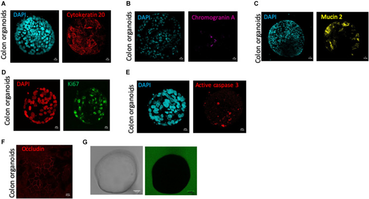
Human colon organoid cell type composition and organoid sealing observation. Colonosphere cross sections are shown for each staining, and all images were acquired with SP8 confocal microscope at 63X objectives and analyzed by Image J software, after 10 days of culture of control organoids. Scale bar 10 μm (A–G). (A) DAPI nucleus (cyan) and cytokeratin 20 (red) staining for colonocytes, (B) DAPI nucleus (cyan) and chromogranin A (pink) staining for enteroendocrine cells, (C) DAPI nucleus (cyan), mucin 2 (yellow) staining for goblet cells, (D) DAPI nucleus (red) and ki67 (green) staining for proliferative cells, (E) DAPI nucleus (cyan) and active caspase 3 (red) staining for apoptotic cells, (F) Occludin staining (red) for tight junction labeling (G) One colonosphere cultivated in the presence of FITC-4 KDa (green) in bright field (left) or fluorescence (right) microscopy.

