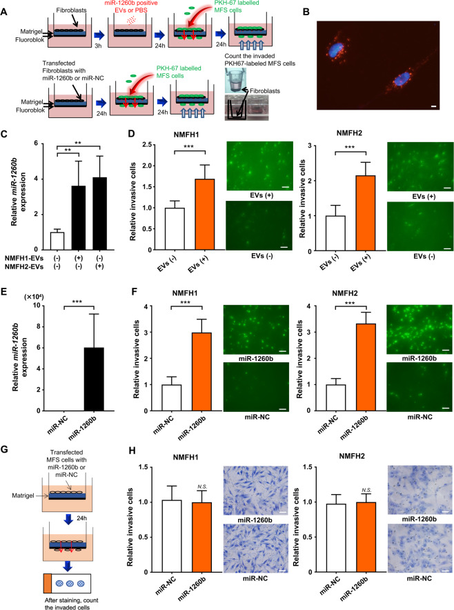
Figure 4.
Tumor infiltration promoted by crosstalk between MFS cells and NFs. (A) Schema of invasion assay using MFS cells and normal fibroblasts educated by tumor-derived EVs or synthetic miR-1260b/control. (B) Fluorescence microscope shows PKH-67-labeled EVs derived from MFS were captured in NFs. Scale bar = 100 nm. (C) Upregulated miR-1260b levels in NFs educated using MFS-derived EVs. **p < 0.01; Student’s t test. (D) Invasion assay. Left, NMFH-1; right, NMFH-2. EV-educated NFs derived from MFS cells promoted cellular invasion of MFS cells. ***p < 0.001; Student’s t test. (E) Upregulated miR-1260b levels in NFs transfected with synthetic miR-1260b mimic or control. ***p < 0.001; Student’s t test. (F) Invasion assay. Left, NMFH-1; right, NMFH-2. NFs upregulated with miR-1260b mimic promoted cellular invasion of MFS cells. ***p < 0.001; Student’s t test. (G) Schema of invasion assay using MFS cells transfected with synthetic miR-1260b or control. (H) Invasion assay. Left, NMFH-1; right, NMFH-2. Scale bar = 100 μm. N.S., not significant; Student’s t test.

