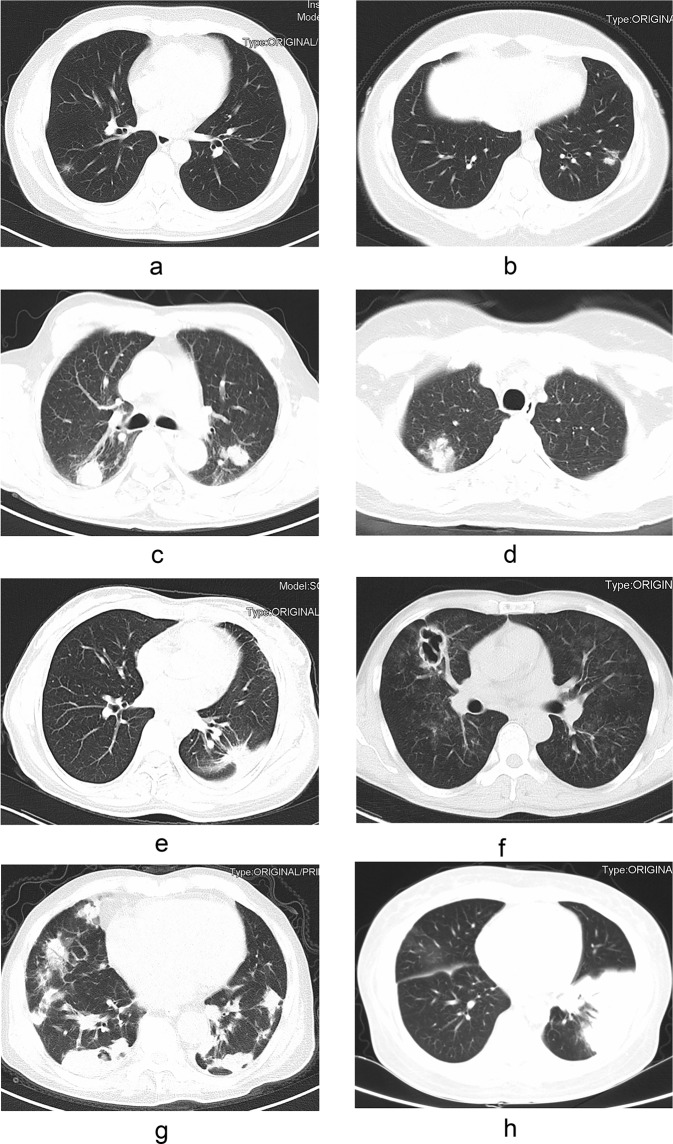
Figure 1.
The findings of chest computed tomography of pulmonary crypotoccosis (immunocompetent patients: (a–e); immunocompromised patients: (f–h): (a) ground glass attenuation; (b) a single small nodule; (c) multiple lung nodules; (d) patchy shadows with air bronchogram; (e) nodular shadow with spiculate boundary; (f) pulmonary cavity; (g) scattered irregular patchy consolidation, nodules and mass shadows in bilateral lungs, and formation of cavities in a few lesions; (h) shadows of miliary nodules in bilateral lungs.

