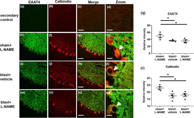
Figure 7.
Reduced EAAT4 expression on cerebellar Purkinje cells was observed in mice exposed to repetitive blast injury. (a) Shows representative images of secondary only controls (no primary antibodies) for Alexa 488, (b) Alexa Cy3, (c) merged, and (d) zoomed (40×). (e) Shows representative immunofluorescent images of EAAT4 (green), (f) calbindin (red), (g) merged, and (h) zoomed images in lobule IX of the cerebellum in control mice at one month after 3X sham and L-NAME administration. (i) Shows representative images of EAAT4, (j) calbindin, (k) merged, and (l) zoomed images at one month after repetitive mTBI. (m) Shows representative images of EAAT4, (n) calbindin, (o) merged, and (p) zoomed images at one month after repetitive mTBI with L-NAME. A significant decrease in (q) EAAT4 (*p ≤ 0.05), and (r) calbindin (*p ≤ 0.05) immunofluorescence was observed in lobule IX of the cerebellum of mice exposed to both repetitive mTBI plus vehicle (*p ≤ 0.05), and repetitive mTBI plus L-NAME (*p ≤ 0.05). One-way ANOVA post hoc Newman-Keuls. Values represent means ± SEM. Arrowheads highlight EAAT4+/calbindin+ Purkinje cell bodies. Scale bars = 30 µm, 20 µm (zoomed).

