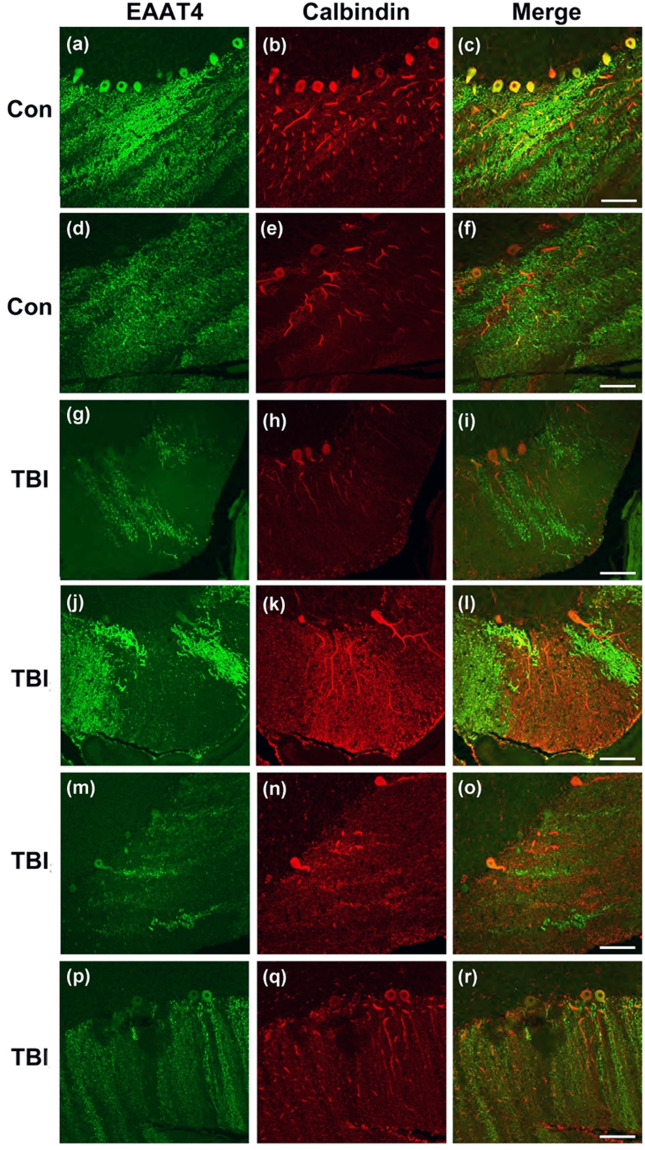
Figure 8.
Dystrophic EAAT4 expression in cerebellar Purkinje cells in human with blast-related mTBI. Confocal microscopy shows representative immunofluorescent images of EAAT4 (green), and calbindin (red) in the cerebella of Veteran/military Servicemember controls with no blast TBI (a–c and d–f, Controls1–2, respectively described in Results), and in the cerebellum of Veteran/military Servicemembers with blast-related mTBI (g–i, j–l, m–o, and p–r, TBIs1–4, respectively as described in Results) Scale bars = 50 µm.

