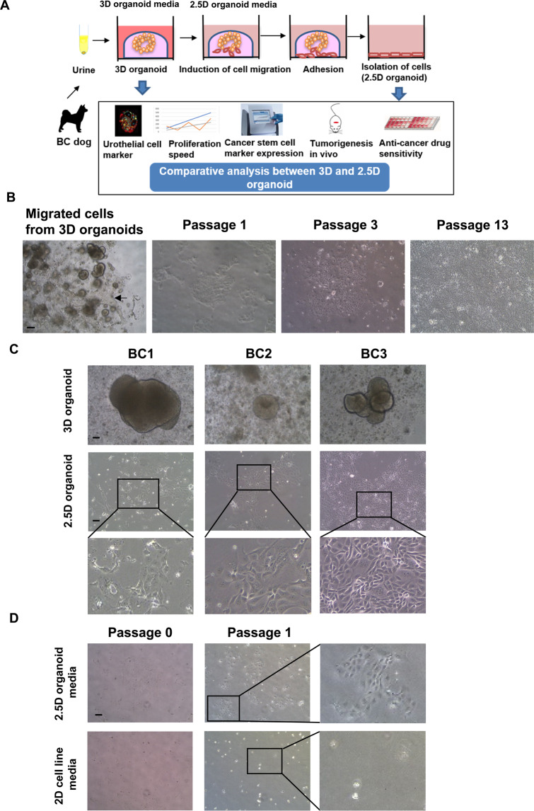
Figure 1.
Generation of dog bladder cancer (BC) 2.5D organoids. Schematic experimental design of a procedure for isolation of 2.5D organoid cells from 3D organoids and the analysis overview (A). Representative bright-field images of the process of generation of 2.5D organoids from 3D ones and serially passaged cells (B). Scale bar: 500 μm. Representative images and enlarged ones (Scale bar: 200 μm) for three different strains of 2.5D organoid cells and their parental 3D organoids (C). Comparison of cell attachment and proliferation between the 2.5D organoid media and 2D cell line media (D). Representative images at passage 0 and 1 and enlarged ones at passage 1 were shown. Scale bar: 500 μm.

