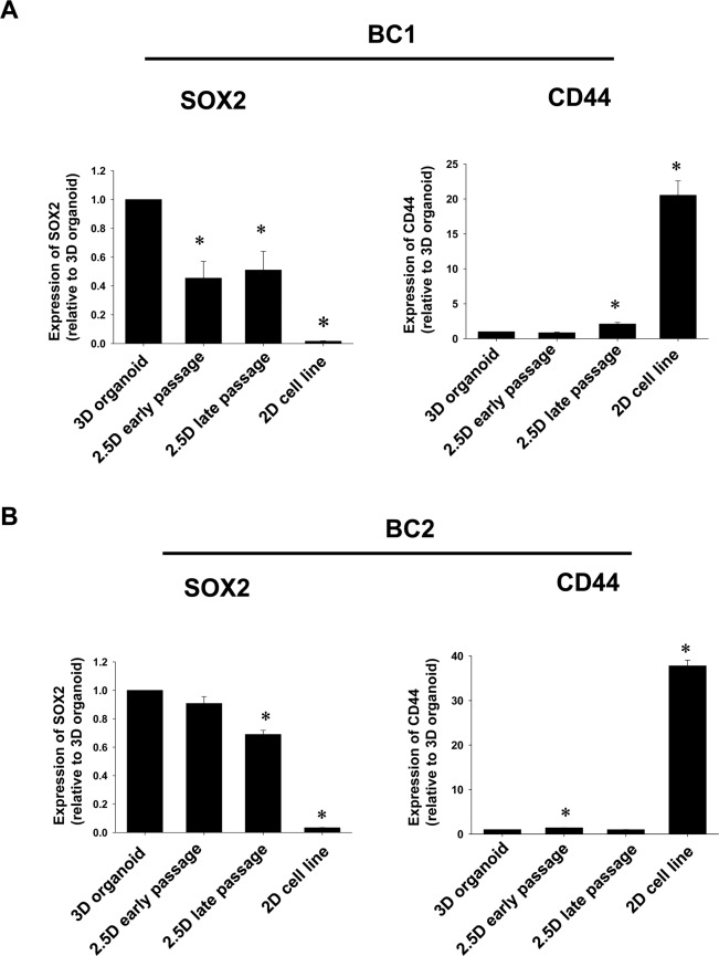
Figure 3.
Comparison of cancer stem cell markers, SOX2 and CD44 between BC 2.5D organoid at the early and late passage and their original BC 3D organoids. Dog urothelial carcinoma cells were used as 2D cell lines. Expression level of SOX2 and CD44 in 3D and 2.5D organoids from different strains (BC1; A and BC2; B) and 2D cell lines was analyzed by quantitative real-time PCR (n = 4) and quantified based on the ratio of expression level to GAPDH. Data were expressed as mean ± SEM. *P < 0.05 vs. 3D organoid.

