Figure 5.
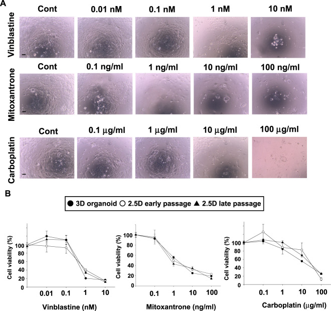
Comparison of the anti-cancer drug sensitivity between BC 2.5D organoids at the early and late passage and their original 3D organoids. After the 2.5D organoids were trypsinized and seeded into 96 well plates, they were treated with vinblastine, mitoxantrone, and carboplatin for 72 h. Representative phase-contrast images of the treated 2.5D organoid cells at early passage were shown (A). Scale bar: 500 μm. Cell viability was assayed by the Prestoblue cell viability kit and 100% represents the cell viability of each control (B, n = 6). Data were presented as mean ± S.E.M.

