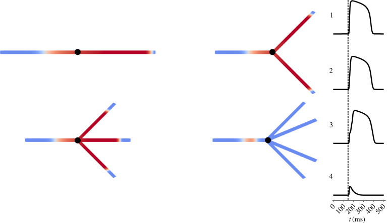
Figure 1.
Tissue fibres with splits inducing variable extents of source/sink mismatch. A schematic demonstration of the meshes used to determine basic electrophysiological properties and the sensitivity to conduction block for different manifestations of ischaemic remodelling. In each case, an initial 2 cm cable allows excitation to reach a steady travelling pulse, which then encounters a split into some number of identical cables, also 2 cm in length. Note that the meshes pictured only represent an illustration of the graph structure, and that in each case the fibre splits into a number of geometrically equivalent fibres despite how the diagrams appear. An example simulation is shown, with failure to propagate through a quadrificated fibre and successful propagation in the other three scenarios. Time courses are plotted at the point marked in black, showing also the delay in activation experienced in the trifurcated fibre. (Online version in colour.)

