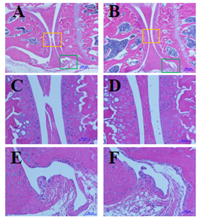
Figure 8.
No differences in histologic features of knee joints between (A, C, and E) human IL8-expressing (hIL8+) and (B, D, and F) control mice. Sagittal sections of knee joints were stained with hematoxylin and eosin; C and D show knee articular cartilage and meniscus tissues, magnified from yellow squares in panels A and B, respectively; E and F show synovial tissues, magnified from green squares in panels A and B, respectively. Scale bars, 200 μm (A and B), 50 μm (C through F).

