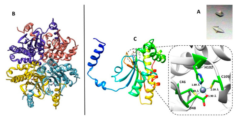
Figure 1.
(A) The shape of BpsβCA crystals, under bright field illumination. (B) Ribbon diagram showing the tetrameric arrangement of BpsβCA. (C) Crystal structure of BpsβCA. Ribbon diagram of the BpsβCA structure, asymmetric unit content, and active site of BpsβCA. The detailing insert shows the enzyme active site with the zinc ion (gray sphere) and its ligands (Cys46, His102, Cys105, and Asp48).

