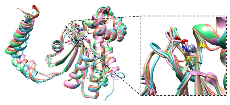
Figure 2.
Superposition of the BpsβCA structure (brown), with the previously determined type II β-CAs from Pseudomonas aeruginosa (cyan, r.m.s.d of 0.784 Å), Porphyridium purpureum (violet r.m.s.d of 0.749 Å), Salmonella typhimurium (green r.m.s.d of 0.785 Å), and Vibrio cholerae (red r.m.s.d of 0.952 Å). The gray sphere represents the zinc atom in the active site. The right panel highlights the active site of type II β-CAs.

