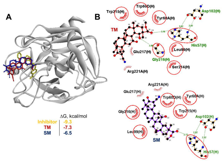
Figure 7.
TM can target thrombin. (A) A stereo presentation of the docked poses of TM and SM in thrombin superimposed on the thrombin inhibitor-bound structure. (B) A 2D representation of the docked poses of TM and SM in thrombin. Common residues, interacting with both the inhibitor and TM, are highlighted in red circles. The comb and green dashed lines represent nonbonding contacts and hydrogen bonds, respectively.

