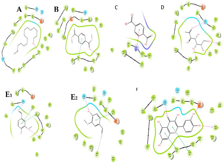
Figure 5.
Interaction and binding of selected test compounds against the C. violaceum CviR protein active site. The negatively charged protein residues are indicated in red, polar residues are in cyan, hydrophobic residues are shown in parrot green, hydrogen interactions (H-bonds) are presented as pink/purple arrows, pi–pi stacking is shown as a green line, and the pi cation as a red line. (A)—Pentadecanol, (B)—Dimethyl terephthalate, (C)—Terephthalic acid, (D)—Methyl mannose, (E1,E2)—Vanillin, and (F)—Quercertin.

