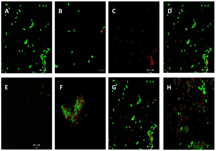
Figure 7.
Observation of P. aeruginosa cells treated and untreated with test extracts of C. aurea ethanol, acetone, and ethyl acetate using confocal laser scanning microscopy (CLSM). (A)—P. aeruginosa ATCC 9721 cells treated 1% DMSO, (B)—Cells treated with C. aurea ethanol extract, (C)—image showing dead cells only treated with C. aurea ethanol extract, and (D)—Combination of live and dead cells treated with C. aurea ethanol extract. (E)—Cells treated with ciprofloxacin (0.06 mg/mL). (F–H) represent cells treated with ethanol, acetone, and ethyl acetate extracts, respectively. Live cells staining appear green, while dead cells show red fluorescence.

