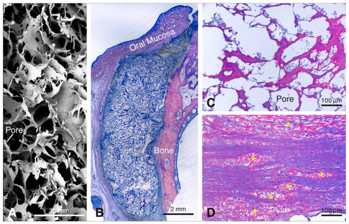
Figure 1.
Scanning electron microscopic image illustrating the three-dimensional-structure of the scaffold (A). Overview of a methylmethacrylate (MMA)-embedded tissue section showing the scaffold between the bone and the soft connective tissue 4 days after implantation (B). The resin section illustrates the pores of the scaffold filled with blood plasma and erythrocytes after 4 days (C). Resin section 90 days after implantation of the scaffold showing elastin fiber packages integrated in the host tissues (D).

