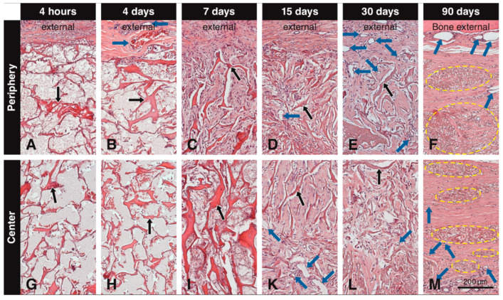
Figure 2.
Tissue sections embedded in paraffin and stained with H&E showing the scaffold (black arrows) at different time points at the periphery and the center. Immediately after implantation and after 4 days, the pores of the scaffold are mainly filled with blood plasma/wound fluid and erythrocytes (A,B,G,H). At 4 days, blood vessels (blue arrows) can be seen adjacent to the scaffold, but not penetrating it (B). At 7 days, cells start to invade the scaffold from the periphery and initial filling of the pores with extracellular matrix begins (C), while these cells do not reach the central portion yet (I). At 15 days, blood vessels are clearly seen in the scaffold pores both at the periphery (D) and in the center (K), while mesenchymal-like cells inside the pores have increased in amounts, and extracellular matrix fill is more advanced (D,K). At 30 days, blood vessels increase in number at the periphery (E) and in the center of the scaffold (L) and newly formed tissue within the pores presents a compact structure throughout the scaffold (E,L). At 90 days, dense packages of residual elastin (yellow circles) are present at the periphery and the center of the scaffold, along with large blood vessels external to the scaffold and smaller blood vessels in the newly formed soft connective tissue around the residual elastin (F,M).

