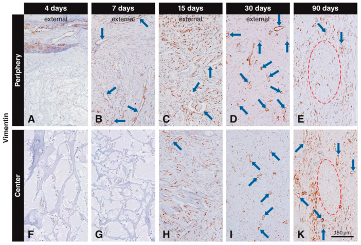
Figure 3.
Immunohistochemical staining for vimentin to label mesenchymal cells. Brown (DAB+) staining indicates vimentin+ mesenchymal cells. At four days, vimentin+ cells are located in the soft connective tissue around the collagen-based scaffold, but have not yet invaded it (A,F). At day 7, vimentin+ cells have invaded the periphery of the scaffold as scattered cells and blood vessels (blue arrows), but have not yet reached the center (B,G). At 15 days, numerous scattered cells and blood vessels positive for vimentin occupy the scaffold pores (C,H). At 30 days, most of the vimentin+ cells are located in the center of the scaffold (I), while some vimentin+ cells can still be found in the peripheral pores and in the external host tissue (D). Labeled cells with an elongated shape can be seen 90 days after implantation in the in the soft connective tissue surrounding the residual scaffold, both at the periphery (E) and in the center (K).

