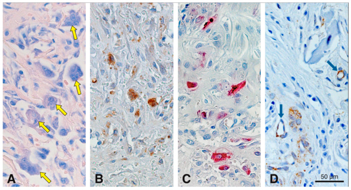
Figure 7.
Histochemical staining for Giemsa and TRAP, and immunohistochemical stainings for cathepsin K and CD86 in serial sections to assess cell-mediated degradation of the scaffold at 15 days in precisely the same region. Medium-sized multinucleated cells (yellow arrows) are differentially stained with Giemsa (A). Cathepsin K (B), TRAP (C) and CD86 (D) stainings reveal that these cells are in direct contact with the collagen-based scaffold. Blue arrows in (D) points blood vessels partially positive for CD86. Cathepsin K+ (B) and CD86+ (D) cells, are stained in brown (DAB+).

