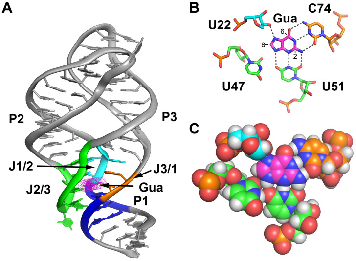
Figure 1.
Structure of the guanine-guanine riboswitch (GR) complex (A) Ribbon cartoon representation of the global structure of the ligand-RNA complex, with key regions in the ligand binding pocket highlighted in color (helix P1, blue; J1/2, cyan; J2/3, green; J3/1, orange). Guanine is shown in magenta. The same coloring scheme is used throughout. This is from PDB 1Y27 [12]. (B) Recognition of guanine by the ligand binding pocket situated in the three-way junction. U51 and C74 form three-hydrogen bond pairing interactions with the guanine ligand. The C8, C6 and C2 positions are denoted on the ligand. (C) The same perspective as in panel B, with a Van der Waals spheres to emphasize the space adjacent to the C8, C6 and C2 positions that serves as the starting point for exploring modifications. Hydrogen atoms have been added to fully illustrate the hydrogen bonding interactions.

