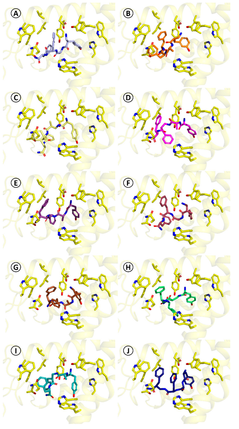
Figure 4.
Binding modes of compounds 8–17 as predicted by docking. The pictures are oriented in the same projection as Figure 2. Only several side-chains of the receptor binding site (yellow sticks) are shown. (A) [(R)-β2hTyr1]-TAPP*, 8, (B) [(R)-β2h-m-Tyr1]-TAPP*, 9, (C) [(S)-β2hAla2]-TAPP, 10, (D) [(R)-β2hPhe3]-TAPP, 11, (E) [(R)-β2hPhe4]-TAPP, 12, (F) [(S)-β2hTyr1]-TAPP*, 13, (G) [(S)-β2h-m-Tyr1]-TAPP*, 14, (H) [(R)-β2hAla2]-TAPP, 15, (I) [(S)-β2hPhe3]-TAPP, 16, (J) [(S)-β2hPhe4]-TAPP, 17.

