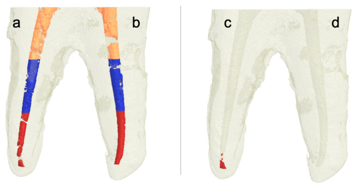
Figure 3.
Three-dimensional rendered images constructed from micro-CT scans of root canals filled with Ca(OH)2. After placement (a,b); and after removal (c) XP/SI; and (d) XP/PUI). Note that the orange color indicates Ca(OH)2 in the coronal third, the blue color indicates Ca(OH)2 in the middle third, and the red color indicates Ca(OH)2 in the apical third.

