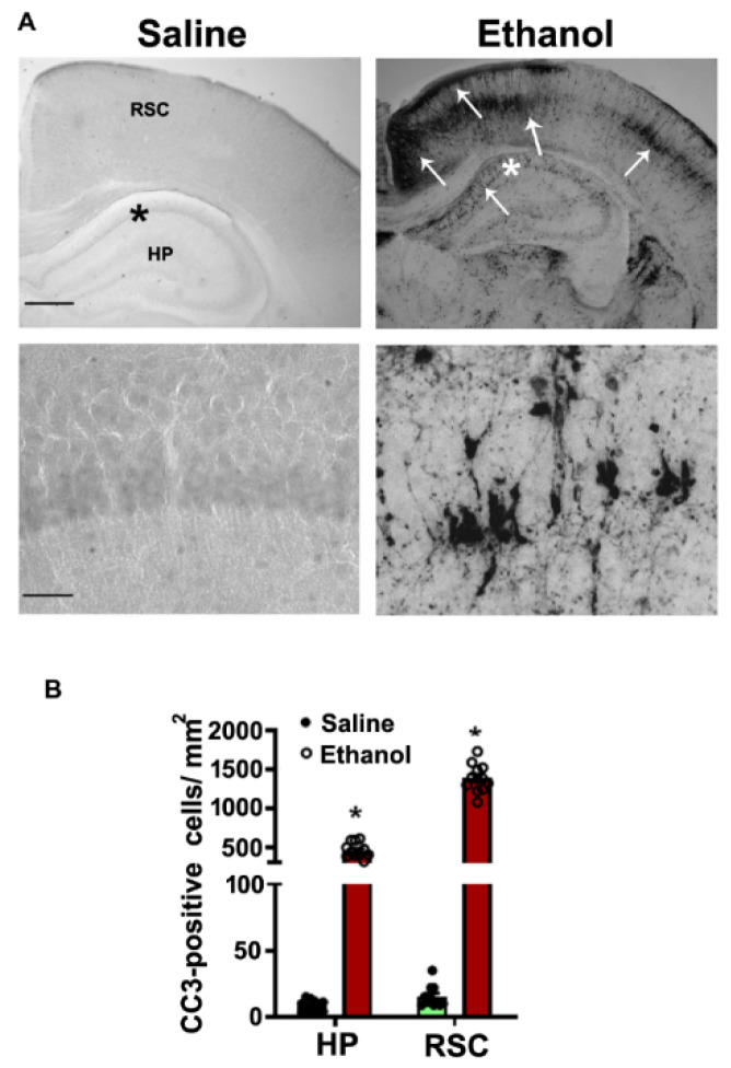
Figure 1.
Enhanced CC3-positive cells in the P7 mouse HPand NC brain regions in response to high-dose ethanol exposure. The free-floating coronal brain sections (HP, and RSC (retrosplenial cortex)) were obtained after saline and 8 h ethanol-exposed mice and sections were subjected to IHC analysis with anti-rabbit-CC3 (A). The arrows indicate the CC3-positive neurons in the HP and RSC. Scale bars = 200 μm. The hippocampal region was enlarged to show the CC3-positive cells (*). CC3-positive cells were counted in the HP and RSC brain regions (B). Error bars, SEM (* p < 0.05 vs. the saline group, n = 6 pups/group).

