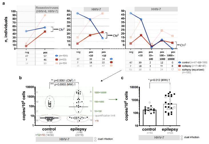
Figure 2.
HHV-7 load in whole peripheral blood (WPB) of the patients with epilepsy (including cases with dual HHV-7 and HHV-6 infection) and control group individuals: (a) comparison of frequency of HHV-7 positive (pos) and negative (neg) cases in epilepsy patients and control individuals; (b) comparison of viral load and frequency of HHV-7 positive epilepsy patients and control individuals, with red ovals are marked low viral load (<10 copies/106 cells) cases, with green ovals—elevated viral load (> 10 copies/106 cells) cases; (c) comparison of elevated viral loads of HHV-7 in epilepsy patients and control individuals; light symbols indicate cases with double HHV-7 and HHV-6 infection (b,c); asterisks in a represent a significance level of differences between groups (** p < 0.01, **** p < 0.0001).

