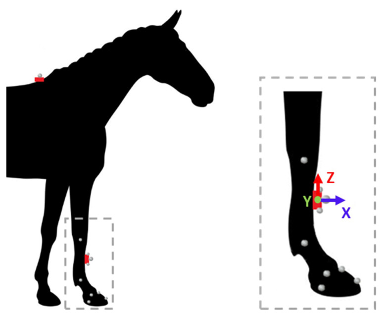
Figure 1.
Placement of the two IMUs (represented in red) and the kinematics markers according to points of interest: six at anatomical points: carpal joint, metacarpo-phalangeal joint, hoof (toe, heel, front coronary band, lateral coronary band), one at the center of wither’s IMU and three on canon bone’s IMU (center, up lateral part, down lateral part). One additional free marker was used for synchronization keystroke on the wither’s marker.

