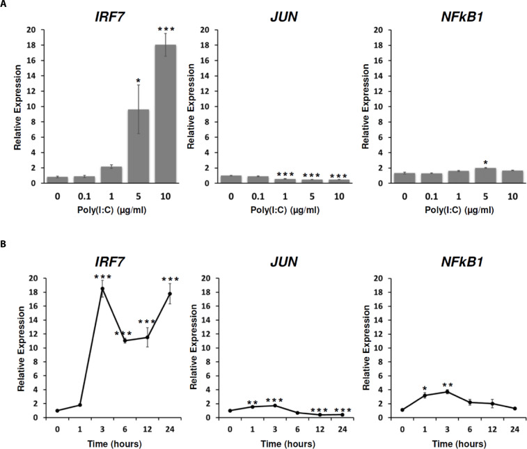
Fig. 3. Dose- and time-dependent expression patterns of TLR signaling-associated transcription factors (TFs) by poly(I:C) treatment.
The expressions of IRF7, JUN, and NF-κB1 in DF-1 cells were analyzed in poly(I:C)-treated conditions with concentrations of 0, 0.1, 1, 5, and 10 µg/mL for 24 h (A) and with concentration of 10 µg/mL for 1, 3, 6, 12, and 24 h (B). The statistical analysis was performed to assess statistical significance between each treated condition and the non-treated control. Error bars were expressed as SEM *p < 0.05, **p < 0.01, ***p < 0.001.

