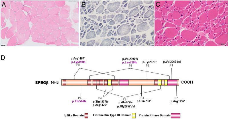
Figure 1:
Histological examination of patients’ muscle biopsies and SPEG schematic. (A) Hematoxylin & eosin (H&E) staining of Patient 1’s muscle biopsy specimen, performed at 9 years of age. The muscle biopsy shows a mild increase in fiber size variability, several atrophic fibers, and only a few internal/central nuclei, consistent with non-CNM CM. (B) SDH and (C) H&E staining of Patient 2’s muscle biopsy, performed at 3 years of age. The muscle biopsy reveals marked variability in fiber size with hypotrophic type 1 fibers and hypertrophic type II fibers with many central nuclei, consistent with CNM. Scale bar 50μm for all images. (D) Schematic of SPEGβ domain organization with positions of identified mutations generated by IBS (Illustrator for Biological Sequences). Mutations affecting both SPEGα and SPEGβ are in black, while mutations affecting only SPEGβ are in pink.

