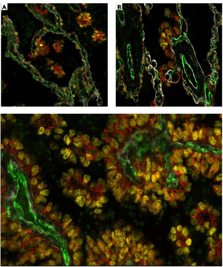
Figure 5: Immunofluorescence (IF) staining:

(A and B) IF stain from an area of lung parenchyma beyond the edge of the tumor (STAS area) shows the nuclear yellow TTF1 staining of the micropapillary (MIP) STAS cells with attachments to the alveolar walls often in areas where the tumor cell cytoplasm is expressing membranous orange staining by E-cadherin (Figure 5A and B). The STAS cells focally are in close apposition to the pre-existing capillaries in the alveolar septa (CD31 positive, green). The alveolar walls show preserved architecture with thin regular capillaries with areas showing preserved type IV collagen of the alveolar wall basement membrane (Collagen IV, white). No endothelial cells or CD31 staining are seen within the STAS clusters. C: IF stain at one level of the main tumor shows the micropapillary (MIP) floret clusters adjacent to papillary structures of the main tumor. The section shows the MIP clusters attached to each other and attached to the papillary structures. The MIP clusters within the air spaces show no CD31 expression indicating lack of a central endothelial lined vascular core (CD31 green negative), differentiating the MIP clusters from the tangential cut of papillary structures.

