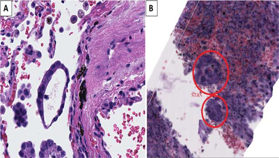
Figure 6:

A) Micropapillary (MIP) STAS appears as a ring-like structure in 2D. (B) however it is seen as a ball-like structure in 3D formed of malignant cells forming a sphere with a wall consisting of a single layer of tumor cells with a central empty lumen (H&E, original magnification x20)
