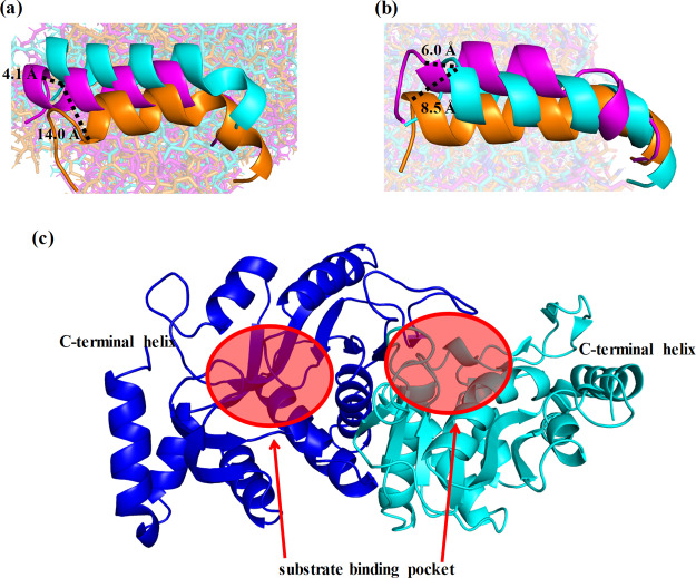
Figure 4.
Differences in the locations and structures of C-terminal helices of dimers. C-terminal helices in (a) chain A and (b) chain B were compared among the wild-type (cyan), middle variant (orange), and Simp-2 (purple). The dotted lines indicate the distance among the Cα atoms of C-terminal residues in the helices. (c) Positions of substrate-binding pockets and C-terminal helices are shown.

