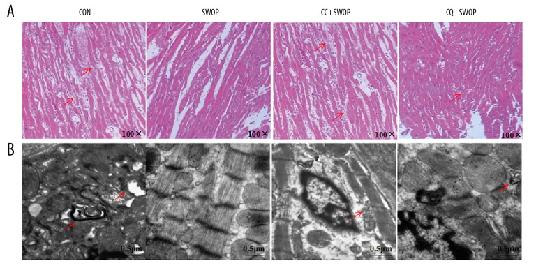
Figure 4.
Sevoflurane attenuated histopathologic organ damage and ultrastructure following I/R. LV tissues were retrieved at the end of reperfusion, and paraffin sections were prepared and subjected to the hematoxylin-eosin (HE) staining. (A) Representative HE staining images are shown (magnification, 100×). (B) LV tissues were harvested for examination of myocardial ultrastructure by transmission electron microscopy. Scale bar, 0.5 μm. The arrowhead indicates that myofilaments were absent or fractured; the nuclear chromatin edge set, and an asterisk indicates mitochondrial edema near the autophagosomes.

