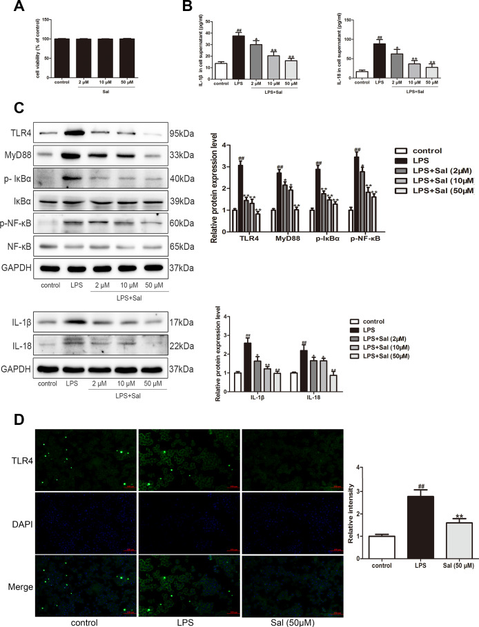
Figure 10.
Sal inhibited LPS-induced BV2 cells inflammation response via TLR4/MyD88/ NF-κB signaling pathways. (A) Effect of different doses of Sal on BV2 cell viability. BV2 cells (1 x 10⁴ cells/well) were exposed to different concentrations of Sal (2, 10, 50 μM) for 24 h. The cell viability of BV2 was measured by CCK8 assay. (B) The levels of IL-1β and IL-18 in the supernatant of BV2 cells were determined by ELISA kits (n = 6). BV2 cell were treated with Sal (2, 10, 50 μM) for 2 h, followed by stimulation with LPS (100 ng/ml) for 24 h. (C) Western blotting was performed to determine the expressions of TLR4, MyD88, p-IкBα, p-NF-кB in LPS-induced BV2 cells. The cells were incubated with Sal (2, 10, 50 μM) for 2 h, followed by stimulation with LPS (100 ng/ml) for 30 min. Western blotting was performed to determine the expression of IL-1β and IL-18 in LPS-induced BV2 cells. The cells were incubated with Sal (2, 10, 50 μM) for 2 h, followed by stimulation with LPS (100 ng/ml) for 24 h. (D) Immunofluorescence staining of TLR4 in BV2 cells. The cells were incubated with Sal (50 μM) for 2 h, followed by stimulation with LPS (100 ng/ml) for 30 min. Original magnification: x200. All data are represented as mean ± SD. # P < 0.05, ## P < 0.01 vs. control group, *P < 0.05, **P < 0.01 vs LPS group.

