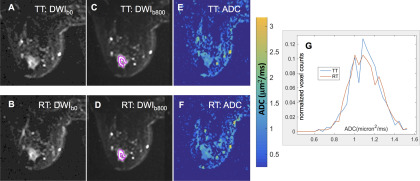
Figure 1.
Sample images and apparent diffusion coefficient (ADC) histograms (bin size, 0.04 μm2/ms) from a typical ACRIN 6698 patient with invasive breast cancer. Grayscale diffusion-weighted imaging (DWI) images for b = 0 s/mm2 (A, B) and b = 800 s/mm2 (C, D) illustrate solid tumor region of interest (ROI) segmentation on 1 slice for (A, C) test (TT) and (B, D) retest (RT) scans. The color images (E, F) show the corresponding ADC maps using the quantitative scale provided in the color bar. Normalized ADC histograms (G) are plotted for the full multislice tumor ROI (red: RT, blue: TT).

