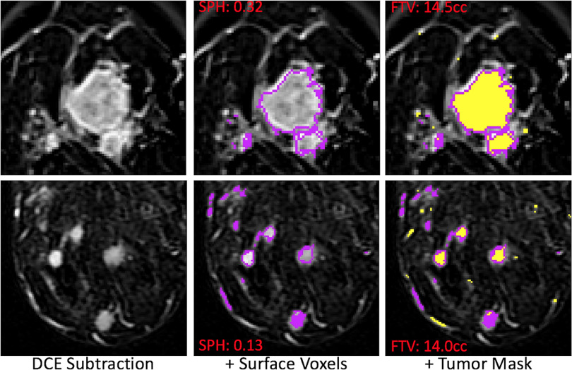
Figure 2.
Example magnetic resonance (MR) images of patients with different morphologic readings. Representative slices of axial magnetic resonance imaging (MRI) were shown from 2 patients. The morphologic readings were 1 and 3 for the top and bottom row, respectively. The sphericity (SPH) values are 0.32 for the top and 0.13 for the bottom. However, the tumor volumes are close (14.5 cc for the top and 14 cc for the bottom).

