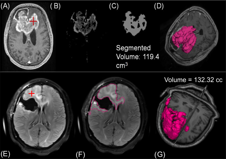
Figure 3.
For the semiautomated CE-T1w segmentation, (A) a clinician must place a seed in the region of interest. (B) Otsu thresholding and filtering identifies extent of enhancement. (C) Indicates the segmented contour for a representative slice after region growing. (D) The entire volume of segmented lesion. The FLAIR segmentation also requires (E) users to make a seed within the hyperintense region. (F) After smoothing, thresholding, and region growing, the contour has segmented the extent of hyperintensity. (G) A volumetric representation of FLAIR hyperintensity for this patient.

