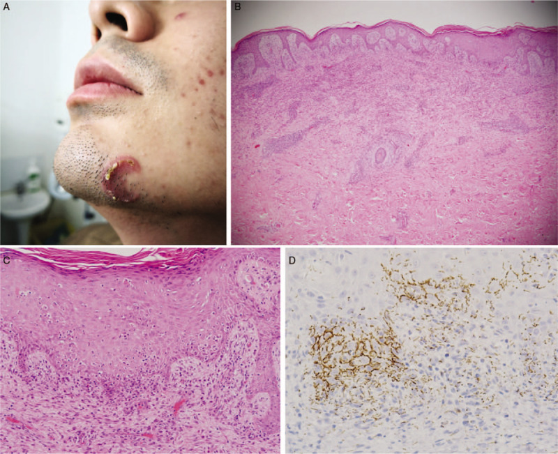
Figure 1.

(A) A solitary annular plaque on the patient's left jaw. (B, C) Section of biopsy showing superficial and deep perivascular inflammatory infiltration with numerous plasma cells, lymphocytes and eosinophils, as well as acanthosis and orthokeratosis with poly-morphonuclear neutrophils exudate in the epidermis (hematoxylin-eosin staining, original magnification ×40, ×200, respectively). (D) Immunohistochemical staining showing numerous spirochetes in the epidermis (original magnification ×400).
