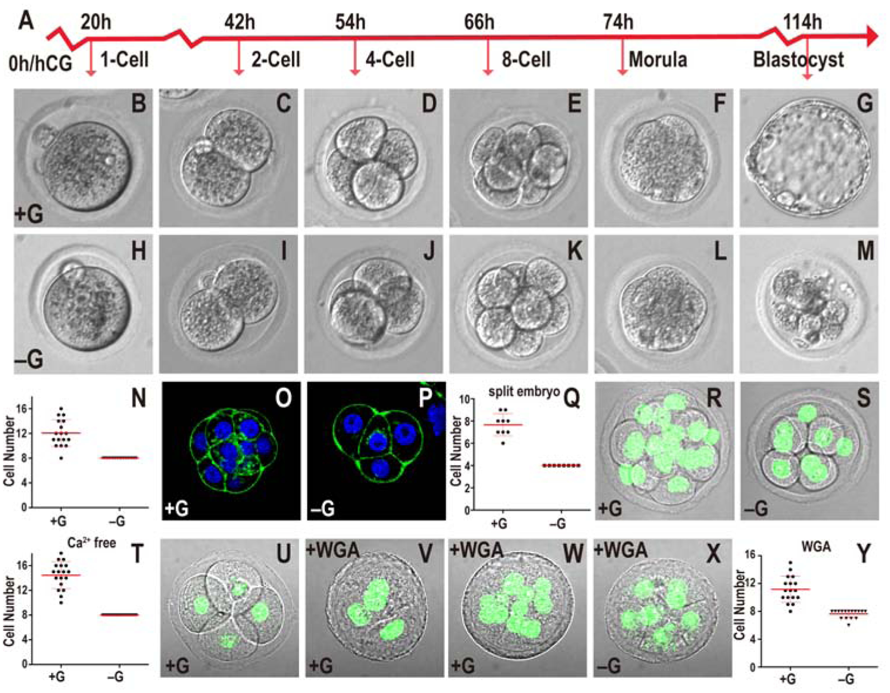
Figure 1. Role of glucose in early embryonic development.

In all figures, hours (h) is following human chorionic gonadotropin (hCG) injection. Growth media including or lacking glucose indicated as +G or −G respectively. Zygotes are isolated at 18h and cultured until the specified hours (h) post hCG. All quantitative data include mean ± SD.
(A) Timeline of preimplantation development. Morula is 78h post hCG that in +G media are at post-compaction 8–16 cell stage with no hint of blastocyst cavity formation
(B-N) Embryos cultured in +G (B-G) or −G (H-M). The times and stages are as indicated in (A). In −G, the embryo fails to make a blastocyst (compare G and M). Every embryo in −G (n=17) is blocked at the 8-cell stage (N).
(O-Q) 2-cell embryos mechanically split into two individual blastomeres and grown in +G (O) or −G (P) until 78h. In both cases the embryos compact at 4-cell. In +G, split embryos proceed to 8-cells but −G embryos are blocked at 4-cell (Q). Note: As 2-cell embryos are split at 46h, they are “4-cell” at 78h.
(R-T) Ca2+ depletion prevents compaction in both +G (R) and −G (S) 78h embryos. Quantitation (T) shows +G embryos contain 10–18 cells (n=18) and −G (n=21) is blocked at 8-cells.
(U-Y) +G grown 4-cell embryos compact when WGA is added (56h; U, 58h; V) proceed to develop beyond 8-cells (n=17) (78h; W, Y). In −G, WGA treated embryos block at 8-cells (n=16) (78h; X, Y).
