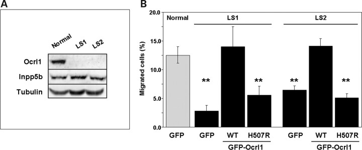
Figure 3.
Dermal fibroblasts from LS patients show cell migration defects linked to the absence of Ocrl1. (A) Whole cell lysates from LS patient and normal control cells were resolved by SDS–PAGE and the presence of Ocrl1 and Inpp5b was investigated by western blotting with specific antibodies. Tubulin was used as a loading control. (B) Normal and LS fibroblasts transfected with the indicated cDNAs were assayed for migration in transwells as described under Materials and Methods. Values represent the mean ± SD of at least three independent determinations. Statistical significance was estimated using pairedt-tests with a correction for significant threshold according to the number of test performed (α = 0.05/3 = 0.017). **P < 0.017.

