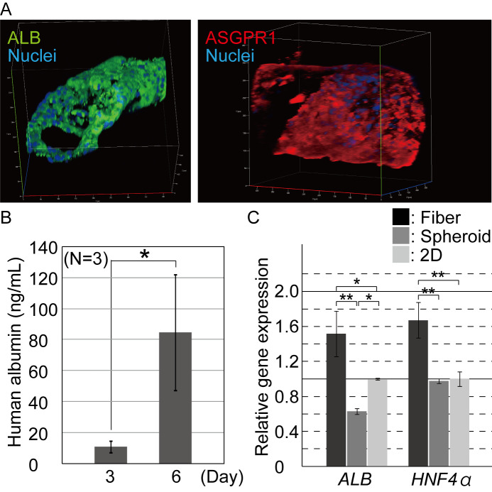
Fig 3. Characterization of the hepatic function of the encapsulated hepatocytes.
(A) Immunocytochemistry was performed for hepatic marker proteins 7 days after hepatocyte cultivation. The iPSC-hepatocytes were positive for albumin and ASGPR1. (B) ELISA was performed for quantifying the albumin secreted into the culture medium. Secreted albumin was detected and its contents increased in the fibers during the culturing process (n = 3, P < 0.07). The error bars represent the standard deviation (s.d.) of triplicate samples. (C.) Quantitative RT-PCR was performed for the hepatic marker genes, ALB, and HNF4α. The expression of the marker genes in the hepatocytes from the cell fiber culture was significantly upregulated, compared to those from the conventional spheroid and 2D culture conditions (n = 3, *; P<0.05), **; P<0.01). The error bars represent the s.d. of triplicate samples.

