
Fig 6. APOE4 exacerbates spreading of αSyn pathology.
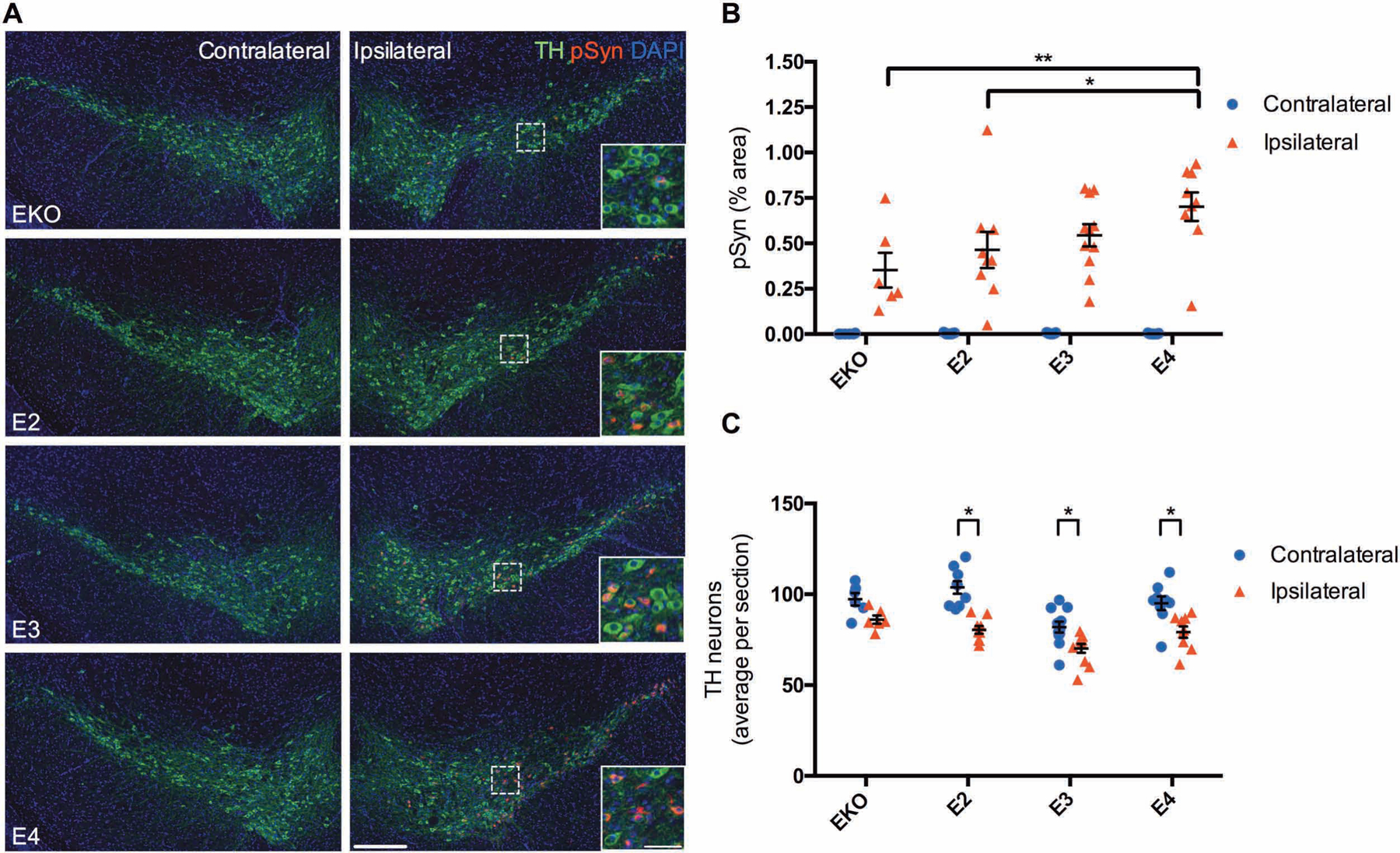
(A) Representative images showing pSyn pathology within the SNpc three months after unilateral injection of αSyn PFFs into the striatum of EKO (n=6), E2 (n=9), E3 (n=11), and E4 (n=9) mice. Scale bar, 250 μm; inset scale bar, 50 μm. (B) Quantitation of the percent area covered by pSyn staining in the SNpc. Data are expressed as mean ± SEM, two-way ANOVA with Tukey’s multiple comparisons test *p<0.05, **p<0.01. (C) Cell counts of TH-positive neurons from 4 sections spaced 150 μm apart. Data are expressed as mean ± SEM, multiple t-tests with correction for multiple comparisons using the Holm-Sidak method.