. 2020 May 11;9:e53789. doi: 10.7554/eLife.53789

Figure 1. HSV-1 pUL7:pUL51 forms a 1:2 heterotrimeric complex in solution.

(A) SEC-MALS analysis of recombinant full-length pUL7:pUL51 complex. Weight-averaged molecular masses (colored solid lines) are shown across the elution profiles (normalized differential refractive index, dRI, colored dashed lines) for samples injected at 2.4, 4.9 and 9.7 mg/mL (blue, orange and purple, respectively). The expected molecular masses for 1:1, 1:2 and 2:4 pUL7:pUL51 complexes are shown as dotted horizontal lines. (B) Averaged SAXS profiles through SEC elution peaks corresponding to 1:2 (blue) and 2:4 (red) complexes of pUL7:pUL51. Fits of representative GASBOR ab initio dummy-residue models to the scattering curves for each complex are shown in yellow. χ2, fit quality. p, Correlation Map (CorMap) P-value of systematic deviations between the model fit and scattering data (61). (C) Refined DAMMIN dummy-atom model reconstruction of the 2:4 pUL7:pUL51 complex, shown in two orthogonal orientations. (D) Representative GASBOR dummy-residue model of the 2:4 pUL7:pUL51 complex, shown in two orthogonal orientations. This model comprises an anti-parallel dimer of heterotrimers, although we note that parallel dimers are also consistent with the scattering data. (E) SEC-MALS of pUL7:pUL51(8-142) complex. Elution profiles and molecular masses are shown as in (A) for recombinant pUL7:pUL51(8–142) injected at 0.6, 1.1 and 3.9 mg/mL (blue, orange and purple, respectively). (F) Schematic representation of pUL7 and pUL51. (G) Averaged SEC-SAXS profile through pUL7:pUL51(8–142) elution peak. Fit of a representative GASBOR ab initio dummy-residue model to the scattering curve is shown in yellow. (H) Refined DAMMIN dummy-atom model reconstruction of pUL7:pUL51(8–142) complex. (I) Representative GASBOR dummy-residue model of pUL7:pUL51(8-142). (J) Plot of the Guinier region (sRg < 1.3) for SAXS profiles shown in (B) and (G). The fit to the Guinier equation (yellow) is linear for each curve, as expected for aggregate-free systems. (K) p(r) vs r profiles for SAXS profiles shown in (B) and (G). (L) Dimensionless Kratky plot of SAXS profiles shown in (B) and (G). The expected maximum of the plot for a compact globular domain that conforms to the Guinier approximation is shown (sRg = √3, (sRg)2I(s)/I0 = 3e−1, grey dotted lines).

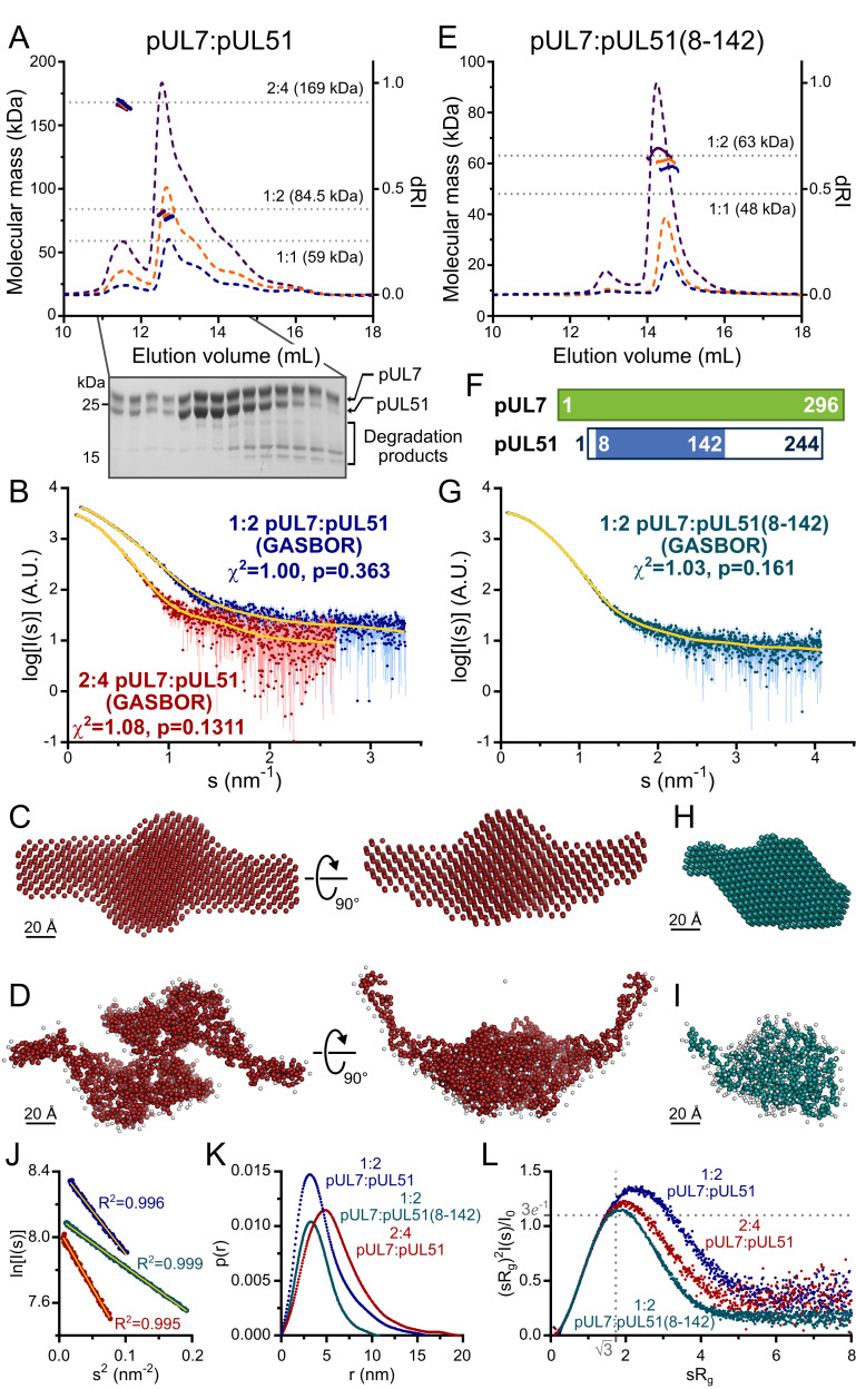
Figure 1.

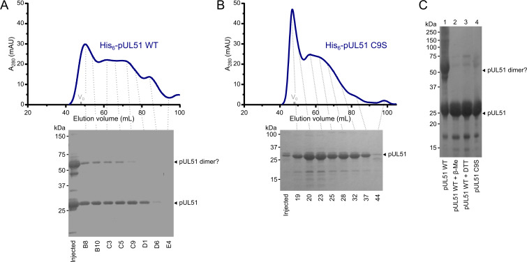
Figure 1—figure supplement 1. HSV-1 pUL51 forms large soluble aggregates when purified in isolation.

Figure 1—figure supplement 1.

(A, B) SEC elution profiles of (A) His6-tagged wild-type pUL51 and (B) His6-tagged pUL51 where Cys9 was substituted with serine (C9S). Proteins were injected onto an S200 16/600 column (GE Healthcare) equilibrated in 20 mM Tris (pH 7.5), 200 mM NaCl, 1 mM DTT. Both proteins have extended elution profiles with peaks near the column void volume (V0), consistent with their forming large soluble aggregates. Coomassie-stained SDS-PAGE analysis of eluted SEC fractions are shown beneath each chromatogram. Note that there is a higher molecular weight band in (A), consistent with the presence of an SDS-resistant pUL51 dimer, despite the presence of 1 mM DTT in the SEC buffer and 2 mM DTT in the SDS-PAGE loading buffer. (C) Purified His6-tagged wild-type pUL51 was subjected to SDS-PAGE either without additional treatment (lane 1) or following incubation with 50 mM β-mercaptoethanol (lane 2) or 50 mM DTT (lane 3). Comparison with the His6-tagged pUL51 C9S mutant (lane 4) confirms that Cys9, the residue that becomes palmitoylated in mammalian cells (Nozawa et al., 2003), mediates disulfide bond mediated dimerization of recombinant wild-type pUL51. C9S substituted pUL51 (or truncations thereof) was thus used for all subsequent experiments with purified proteins.
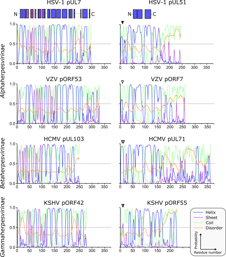
Figure 1—figure supplement 2. Predicted secondary structure of pUL7 and pUL51 homologues from representative human α-, β- and γ-herpesviruses.

Figure 1—figure supplement 2.

Analyses of amino acid sequences were performed as described in Materials and Methods. Per-residue probabilities of forming α-helix (blue), β-sheet (purple) or coil (green) are shown, as is the probability of disorder (orange). Residues that are known (solid triangles) or predicted (empty triangles) to be palmitoylated are marked: Cys9 of HSV-1 pUL51, Cys9 of VZV pORF7, Cys8 and Cys13 of HCMV pUL71, Cys10 and Cys11 of KSHV pORF55. Regions of pUL7 and pUL51 α-helix and β-sheet observed in the pUL7:pUL51(8–142) core heterodimer structure are shown above the predictions as boxes. The predicted pUL7 and pUL51 secondary structural elements are largely conserved across herpesvirus families, although the first two helices of pUL7 are not conserved in β-herpesviruses like HCMV. Additionally, the C-terminal regions of pUL51 homologues vary in length, although in all herpesvirus families they are predicted to be largely unstructured.