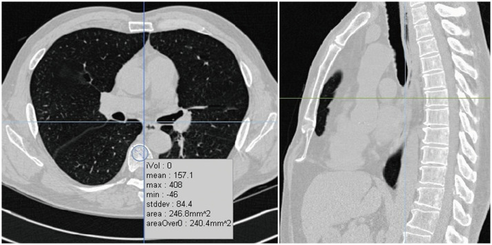
Fig. 1. Measurement of DThorax.
After inspecting sagittal and coronal reconstructed images, central section of axial CT images at each vertebral level were selected. In this selected slice, round region of interest as large as possible was manually set to encompass anterior portion of each vertebral body. Cortical bone area, with large veins, as well as calcified herniated disks, were excluded from region of interest using manual free tracing. DThorax = thoracic vertebral bone density measured on chest CT

