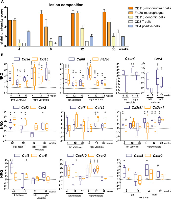
Fig. 6.
Profiling the immune response from the acute to the chronic stage of cardiomyopathy in Dsg2MT mice reveals persistent upregulation of macrophages together with dendritic cells and T cells as well as phase-specific chemokine/chemokine receptor upregulation. a The histogram depicts the results of a semiquantitative microscopical intensity score assessment of immunohistochemical immune cell detection in myocardial scars of Dsg2MT mice (n = 3–5 for each age group). b Tukey's whisker plots show the results of qRT-PCR (normalized relative quantification; NRQ) performed on RNA isolates from right and left ventricles of Dsg2MT and wild-type controls (dotted lines). Expression was assessed by using non-parametric Mann Whitney tests: *p < 0.05, **p < 0.01; §p > 0.05 and < 0.06. For details see Supplementary Table 6 and 7. The increase of Cd45 (immune cells), Cd68 and F4/80 (monocytes/macrophages), and Cd3e (T cells) mRNA supports the semiquantitative immunohistochemical results in a. Furthermore, the mRNA expression data suggest that the immune response is more pronounced in the right ventricle than in the left ventricle

