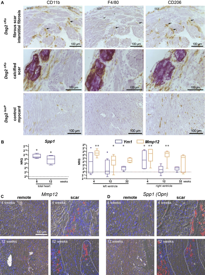
Fig. 7.
Macrophage distribution and macrophage marker expression is lesion type-specific. a depicts the immunolocalization of the macrophage markers CD11b, F4/80 and CD206 in serial cardiac sections (from left to right) of Dsg2cKO mice with interstitial fibrosis and fibrotic or calcified scars and of Dsg2loxP control mice at 35 weeks (n = 3 and n = 2, respectively). SEF marks a subepicardial fibrotic scar, arrows point to areas with interstitial fibrosis. b Tukey's whisker plots show the quantification (normalized relative quantification; NRQ) of RNAs encoding the macrophage markers Spp1, Ym1, and Mmp12 in Dsg2MT and wild-type control mice (the dotted line indicating relative mean levels of the wildtype). Mann Whitney tests were applied to compare Dsg2WT and Dsg2MT mice at each time point: *p < 0.05, **p < 0.01. For more details see Supplementary Table 7. c Microscopy of in situ hybridizations detecting Mmp12 and Spp1 mRNA (red signal) and nuclei (blue) in paraffin sections of Dsg2cKO or Dsg2MT and corresponding control hearts at the age of 4 and 12 weeks. Sections of 3–4 Dsg2MT/cKO and 2–3 Dsg2WT/loxP hearts were assessed for each age group. Lesions are demarcated by broken lines

