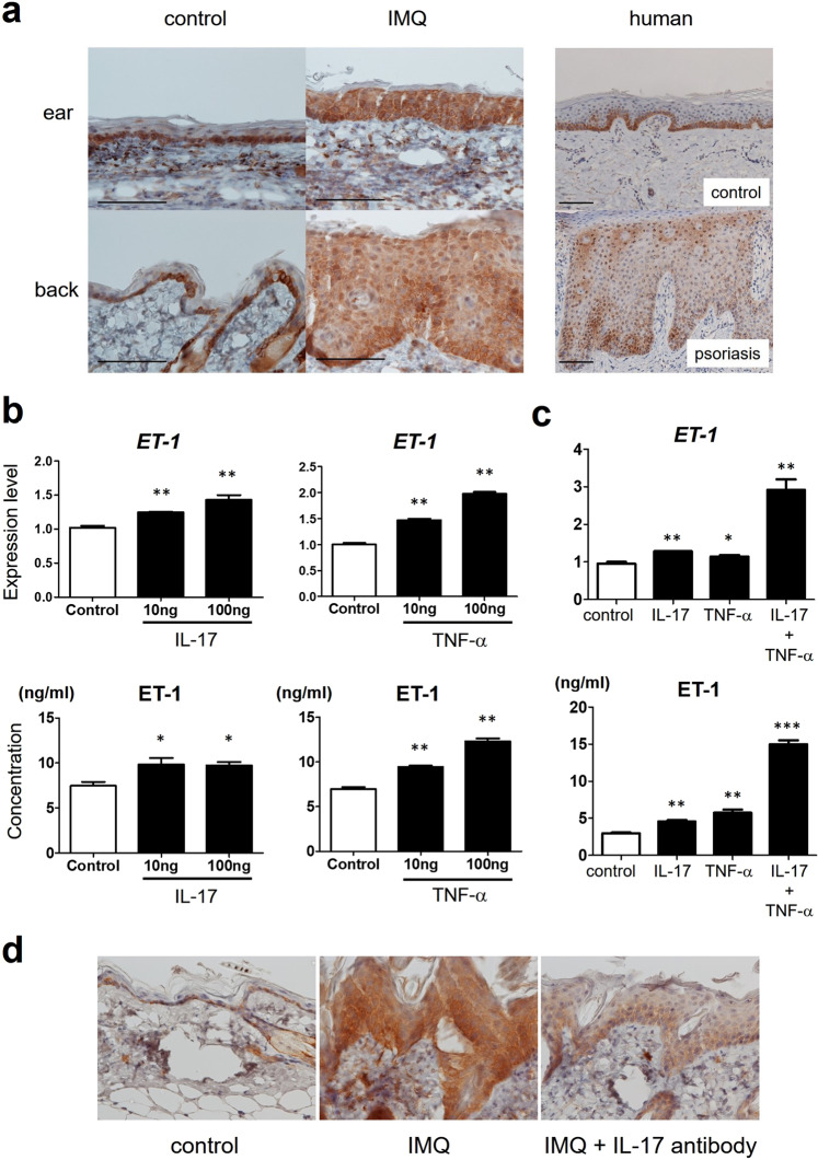
Figure 1.
The expression of ET-1 in mouse and human psoriasis. (a) Immunohistochemical staining for ET-1 in normal skin and psoriasis. Expression of ET-1 was preferentially confined to basal keratinocytes in control mouse or normal human skin (n = 5). In IMQ-induced murine psoriasiform dermatitis (n = 5) or human psoriasis (n = 5), ET-1 expression was detected widely in the whole epidermis. Scale bar: 50 μm. (b) NHEKs were cultured with or without IL‐17 or TNF-α for 24 h. (c) NHEKs were cultured with or without IL‐17, TNF-α, or both for 24 h. Expression levels of mRNA of ET-1 in NHEKs were determined using quantitative PCR. Concentrations of released ET-1 were also measured in cell-free supernatants by ELISA. Data are shown as mean ± SEM. Results are representative of similar results obtained in three independent experiments. *P < 0.05, **P < 0.01 versus the control group (without IL-17 or TNF-α treatment). (d) ET-1 expression in psoriatic epidermis after local application of IL-17 neutralizing antibody. Mice were applied topical IMQ cream daily for five days. At day 1 and day 4, mice were administered IL-17 neutralizing antibody (150 μg/40 μl). Samples from the back at day 6 from control mice (n = 2), IMQ-treated mice (n = 2), and IMQ-treated mice with IL-17 neutralizing antibody (n = 2) were stained for ET-1.

