Figure 3.
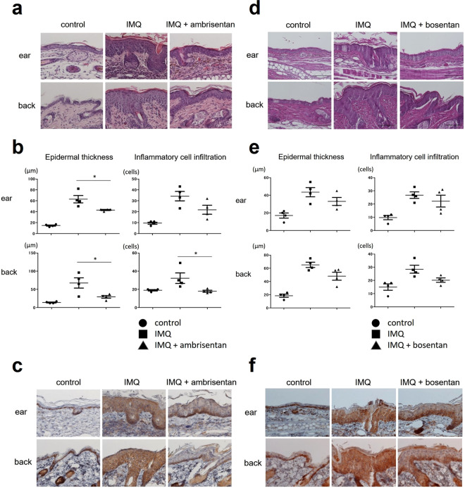
Histological presentation of control skin, IMQ-induced psoriasiform dermatitis, and IMQ-induced psoriasiform dermatitis treated with ambrisentan or bosentan. (a,d) H&E staining of skin tissue harvested at day 6 (×400). Samples from the ear and back of control mice, IMQ-treated mice, and IMQ+ ambrisentan or bosentan-treated mice at day 6 (after treatment for five consecutive days with IMQ) were stained using H&E. (b,e) Epidermal thickness and inflammatory cell infiltration of control skin, IMQ-induced psoriasiform dermatitis, and IMQ-induced murine psoriasiform dermatitis treated with ambrisentan or bosentan. The epidermal thickness and number of dermal inflammatory cells were determined per high-power field from four mice per group. Data are presented as mean ± SEM. *P < 0.05, versus IMQ-treated group. (c,f) Immunohistochemical staining for ET-1 of samples from the ear and back of control mice, IMQ-treated mice, and IMQ+ ambrisentan or bosentan-treated mice (×400). Results are representative of similar results obtained in three independent experiments.

