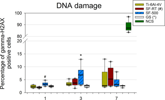
Figure 10.

Flow cytometry of the marker of DNA double-strand breaks gamma-H2AX (phosphorylated histone H2AX) in U-2 OS cells grown for 1, 3 and 7 days on bare Ti-6Al-4V samples (Ti-6Al-4V), on samples coated with as-synthesized silicalite-1 films (SF-RT) or with silicalite-1 films heated up to 500 °C (SF-500), and on the control microscopic glass coverslips (GS). The box plot bold black central line shows the median, and its outer edges represent the 1st and 3rd quartile. *Indicates a significant difference from GS, and # shows the difference from SF-RT with p ≤ 0.05.
