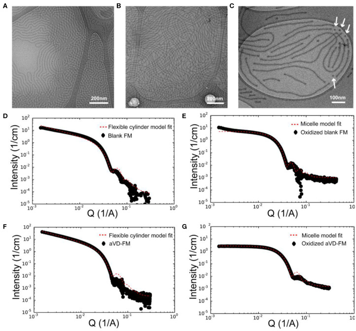
Figure 2.
Characterization of aVD-loaded filomicelles. CryoTEM images of filomicelles (FM) (A) and aVD-loaded FM (B,C). The white arrows identify the morphological transition from filaments to spherical micelles. Small angle X-ray scattering (SAXS) results are shown for blank FM (D,E) and aVD-loaded FM (F,G) in PBS solution (D,F) or 500 mM H2O2 oxidizer (E,G).

