
FIGURE 4.
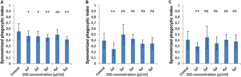
The symmetrized phagocytic index varies according to the DID concentration used for MH-S cell labeling during phagocytosis of unlabeled spores of L. corymbifera and A. fumigatus phagocytosed by DID-stained MH-S cells. (A–C) are as in Figure 2. Three independent biological replicates were carried out. Statistical difference was always measured in comparison to the control experiment and was described as: “ns” means there is no significant difference, *p < 0.05, **p < 0.01, ***p < 0.001. The data represent mean ± standard deviation.